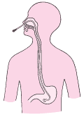
nasogastric tube and / or orogastric DEFINITION
technique of introducing a flexible tube (silicone, polyurethane or reflón) in the patient's stomach through the nose (nasogastric tube) or mouth (orogastric).
OBJECTIVE
-
Administration of enteral feeding.
-
medication administration. Make
-
gastric lavage.
-
Aspirate gastric contents to:
- prevent aspiration in intubated patients or decreased level of consciousness.
- decompress or remove air or fluid from the stomach.
- prevent suture failure in patients undergoing gastric resection.
- monitoring the progress of HDA.
- analysis of gastric contents in the laboratory.
CONSIDERATIONS CONTRAINDICATIONS
-
If basilar skull fracture, broken facial bones and nasal packing is contraindicated inserting the tube through the nose. In these cases be used via orogastric.
-
In patients undergoing esophageal surgery or stomach, keep caution.
-
If the patient has a deviated septum and nasal disorder, which prevents the tube being in this way, put it through the mouth after removal of dental prosthesis and / or piercing.
EQUIPMENT AND MATERIAL
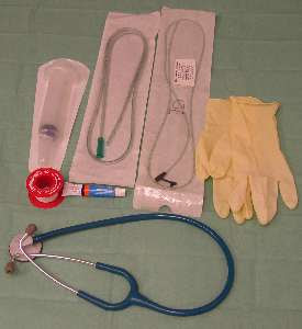
1) Human Resources
2) Material resources
- Nasogastric
. ( consider in their choice: Outside diameter or size and length ).
-
clean gloves
-
Gauze
-
soluble lubricant
-
Lantern
-
Tongue depressor
-
syringe feeding
Stethoscope -
-
hypoallergenic tape or specific material fixing probe
-
drainage bag
- Vacuum
-
Guedel cannula insertions oral
-
Maguilla Clips oral insertions
-
pH test strips to determine
-
Water Tumbler
PROCEDURE
1) Preparation of the patient:
-
If the patient is conscious and capable of understanding, explain the procedure. In the case of newborns or infants, report the procedure to the family. Background: technique is very annoying and difficult to achieve without the cooperation of the patient. It can also cause anxiety in the family if they do not know the procedure.
-
Inspect nasal and oral cavity of the patient. If the patient is conscious ask you to relax and breathe normally while cover one nostril, repeat with the other hole. Choose one, through which a greater flow of air. If unconscious, exploring the nostrils with a flashlight looking for irritation, obstruction and / or deformity (eg, nasal septal deviation). Background: The probe will pass more easily through the hole nasal more permeable.
-
Palpate the patient's abdomen. Background: know the degree of abdominal distension prior to assessing the progress going.
-
In case of existing dentures or piercing, removal therefrom. Background: Avoid travel during insertion of the NGA.
-
patient positioning. generally place it in high Fowler's position with pillows behind her head and shoulders. In most cases it will require auxiliary aid to hold the position. But in case of poisoning it will trendelemburg left lateral decubitus. Background: It facilitates the ability to swallow and the force of gravity helps to pass the probe. In the poisonings, the indicated position prevents the toxic pass into the duodenum.
2) Preparation of material
- Choose the diameter of the probe suitable for the patient. Background: Comes measured in French. 1 French = 0.33 mm.
-
Hand washing and glove positioning: The protocol for washing hands is as follows: 1 .- rub our palms vigorously; 2 .- rub our right palm on the back of the left and vice versa; 3 .- rub both palms fingers intertwined, 4 .- rub our backs bent fingers for each hand, rubbing my thumb 5 .- right to left and vice versa; 6 .- rubbing the fingertips of one hand on the palm of the other. Background: Reduces transmission of microorganisms.
-
determine the length of tube to be introduced. 
For this there are two methods: a) distance between the tip of the nose to the earlobe and then to the xiphoid process in case of SNG. If OG would the distance between the mouth, and the xiphoid process through the earlobe; b) Method Hanson: first mark a point 50 cm from the probe, then performed the traditional method. Should insert the probe to the midpoint between 50 cm and traditional brand.
Background: The length is different for each patient.
-
Mark the length obtained with a tape or marker. Background: Avoid measurement errors.
-
Prepare the type of attachment of the probe. If taped, split longitudinally in half. Background: Set the probe and prevent accidental removal.
-
Roll the end of the probe around the hand. Background: Help insertion and decreases the stiffness of the tube. If you want to get more flexibility you can insert the tube in warm water. If we want is to be introduced more rigid in cold water or ice.
-
Lubricate the tube with water soluble lubricant. Background: decreases the friction of the probe to the nasal mucosa. As the water-soluble lubricant should be dissolved accidental insertion of the probe into the lung.
-
Prepare the syringe and stethoscope.
3) Development of technique
-
Stand on the right side if right handed or left if left handed. Background: facilitates the manipulation of the probe.
-
Insert the probe through the hole selected. If the patient working, ask him to hyper extend the neck to gently insert the tube through the nostril floor down and the ear on that side. Background: reduces the discomfort produced by the friction of the probe against the turbinates.
-
You will notice a slight resistance. Apply a slight downward pressure to advance the probe, if not rotate forward SNG and if the resistance still persists, do not twist and remove the probe. Background: If you force the introduction, it can damage the nasal mucous membranes.
-
Flexing the patient's head to his chest and let the patient relax for a moment. Background: step is facilitated to the posterior pharynx as the glottis closes, reducing the possibility that the probe penetrates trachea.
-
Encourage the patient to swallow, giving small sips of water if not contraindicated. If so, asked to swallow saliva. Go to advance the catheter as the patient swallows. Background: Al swallowing facilitates the advancement of the probe.
-
If there is cough, dyspnea, cyanosis, remove the probe. Background: will have been introduced accidentally in the windpipe.
-
repeated if nausea and SNG does not move with swallowing, inspect the throat with the depressor and flashlight. Background: The probe may be in the throat and stimulate the gag reflex.
4) Check the positioning of the probe
-
If the patient is conscious ask you to speak. Background: If the probe has passed through the vocal cords, patient can not talk.
-
Aspirate gastric contents, evaluating color. Background: The color green is often cloudy. It can also be white or brown.
-
Measure the pH of the aspirate. Background: pH of gastric contents is 4 or less, pH of intestinal secretions is 7.5 to 8 and the pH of lung fluid is around 7.6.
-
Breathe air through the tube, placing the stethoscope in the epigastrium. The amount of air breathed vary with the age of the patient. Background: will hear a hissing or gurgling noise. If not, may be in the esophagus, trachea or bronchus. Not a reliable method of verification.
-
Place the end of the NGA in a glass of water. Background: If bubbles is that it is placed in the bronchial tree. Check
-
radiation.
-
Clean and dry the patient's nose. Background: For the fixation does not loosen.
-
Set the probe with a strip of tape or dressing appropriate depending on the patient's age and the area of \u200b\u200bintroducing it. If the patient has skin lesions or burns, hold the tube using tape or band. If the patient is agitated, fix probe also behind the ear. The probe can also be attached to the endotracheal tube. Background: not be fixed in the front, it can cause pressure ulcers on the nose.
-
Keep the patient with the chest elevated 30 to 45 degrees. Background: prevent aspiration pneumonia.
-
Connect the end of the tube drainage, aspiration, nutrition or tweezers.
-
Remove gloves and wash hands.
nursing registry:
This register should note the type of probe, the number of probes, the brand and the possible complications during the technique.
Complications:
-
Erosion of the nasal mucosa, epistaxis.
-
gastric mucosal erosion, gastric hemorrhage.
-
Aspiration pneumonia. Hyperventilation
-
by increased anxiety in conscious patients. Bradycardia
-
by vagal stimulation.
-
hypokalaemia or metabolic alkalosis due to loss of electrolytes if gastric drainage is abundant.
-
obstruction of the probe.
-
esophageal erosion.
-
reflux esophagitis.
-
Pressure sores. Nausea
-
catheter removal
Definition:
Removing a naso-OG, or because it has blocked or does not necessary .
Objective: Elimination
of that path.
Equipment and material:
Human Resources: Nurse
Material Resources: Towel
-
-
- nonsterile gauze non sterile gloves or forceps
- cap probe
Procedure:
- Explain the procedure to the patient.
- Wash hands and wear gloves non-sterile.
- Place patient in semi-Fowler position. Background: Avoid content aspiration stomach.
- at the right side of the patient if they are right or left side if left handed. Background: allows easier handling of the probe.
-
Disconnect the probe from the aspiration or drainage bag. Clamp the probe. Background: Preventing gastric contents remaining in the drain tube to remove it and enter the airway. Remove
-
fixing probe. If the patient is conscious ask you to take a deep breath and pull the probe gently and quickly during patient expiration. However, if the patient is intubated, make sure the correct pressure neumotaponamiento before removal. Background: relaxes the throat and reduces irritation and danger of aspiration. Perform
- hygiene nostrils and mouth.
- Remove gloves and wash hands.
nursing registry:
Register the procedure and if there were complications during the test.
Complications:
- Aspiration of liquid in the probe if it is badly pinched.
- abdominal distension and / or vomiting has not resumed if the intestinal transit.
ANNEX: Age caliber: gastric tube for washing or draining:
- Neonates and Infants (up to 18 months) 5-8
French - -7 years 18 months 8-10
French - 7 years -10 years French 10-14
- 11 years - 14 years 12-16
French gastric tube for enteral nutrition:
Depending on the patient's age, we use a gauge that range between 5-12 French, taking care to use the smallest possible.
SOURCE: http://www.eccpn.aibarra.org/temario/seccion6/capitulo101/capitulo101.htm


